


The thoracic form of osteochondrosis is characterized by degenerative lesions in the intervertebral cartilage and secondary changes in the thoracic vertebrae. Diagnosing the disease is sometimes quite difficult because it is often "masked" like other diseases: myocardial infarction, angina, gastrointestinal diseases.
Characteristics of thoracic osteonecrosis
This type of disease is quite rare compared to cervical and lumbar.
The cause lies in the specific anatomical structure of the chest area:
- it is the longest (consists of 12 vertebrae);
- In this area there is a slight natural bend - physiological kyphosis, which partially relieves the load caused by walking straight;
- The thoracic region articulates with the ribs and sternum, performs the functions of the physiological frame and takes on the main load;
- In cross-section, the thoracic spinal canal has the smallest size;
- Thoracic vertebrae are thinner and smaller in size but have long vertebrae.
Due to these factors, the thoracic part is not particularly mobile, so osteonecrosis in this part of the spine is rare, but its symptoms are quite pronounced: they are quite strong and unpleasant pain associated with the cord. Spinal nerves are compressed, causing irritation in the shoulder. Lumbar organs and upper limbs are located in the abdominal and thoracic cavities. For the same reasons, the manifestations of thoracic osteoarthritis are often atypical, which significantly complicates the diagnosis of pathology and subsequent treatment.
The narrowness of the spinal canal, physiological kyphosis and the relatively small size of the vertebrae create the most favorable conditions for the formation of disc herniation. Because a significant portion of the load mainly falls on the anterior and lateral parts of the vertebral body and intervertebral disc, the intervertebral disc will deflect backward and form a disc herniation, also known as Schmorl's hernia.
The front part of the vertebra is under greater pressure than the back part. For this reason, the development of bone spurs and disc prolapse often occurs outside the spine and does not affect the spinal cord.
Stages of thoracic osteonecrosis
Manifestations of thoracic osteochondrosis are determined by the changes that occur in the intervertebral discs and vertebrae, depending on which four main stages of the disease are distinguished:
- Stage I is characterized by dehydration of the intervertebral discs, as a result of which they lose elasticity and stiffness but retain their normal load-bearing capacity. The process of flattening of the disc begins, its height decreases and protrusions are formed. The pain at this stage is mild.
- In stage II, cracks form in the annulus and record instability of the entire segment. The pain becomes more intense and intense when bending over and performing certain other movements.
- A characteristic sign of stage III is the rupture of the annulus fibrosus and the beginning of the formation of a herniated disc.
- During the transition to stage IV, because the disc no longer has resistance, the vertebrae begin to move closer together, causing spondylosis (disorder of the joints between the vertebrae) and spondylolisthesis. vertebrae (twisting or shifting of the vertebrae). The mobilization of compensatory forces to reduce the load leads to the vertebrae growing, increasing in area, and collapsing. The affected part of the annulus fibrosus begins to be replaced by bone tissue, which significantly limits the mobility of this part.
Degrees of osteochondrosis of the chest
Today, many experts use a different principle of classification, according to which osteochondrosis of the thoracic spine is distinguished not by stage, but by degree with their characteristic features.
How does level one disease manifest? As a rule, it is diagnosed when the disc ruptures due to excessive exertion or sudden movements. In this case, a sharp pain suddenly appears in the spine. Patients compare it to an electric current running through the spine. This condition is accompanied by stress reflexes of all muscles.
The second degree of thoracic osteochondrosis is referred to in cases where instability of the spine appears and symptoms of protrusion of the intervertebral disc develop. This condition is very rare, occurs in episodes of exacerbation and subsequent remission, and is only detected during a thorough diagnostic examination.
What symptoms appear in stage three disease? The pain becomes constant, spreads along the damaged nerve and is accompanied by partial loss of sensation in the upper or lower limbs, changes in gait and severe headaches. At this stage, people often experience difficulty breathing and normal heart rhythm disturbances.
We can talk about moving to level 4 when the symptoms of the disease subside while symptoms of spinal instability still exist (sliding, twisting of the vertebrae, fixation in relation to each other). Bone spurs begin to develop, gradually compressing spinal nerves and compressing the spinal cord.
Typical symptoms and signs
Osteonecrosis in the thoracic region has quite characteristic signs, on that basis it is very possible to diagnose this disease:
- Intercostal neuralgia - pain is often localized in one area, then quickly spreads to the entire chest, forcing the patient to take a certain position and causing significant difficulty in breathing.
- When turning your body, moving your neck, bending, raising your arms, breathing (inhale-exhale), the pain becomes much more intense.
- The muscles in the middle and upper back are severely spasmed. Contraction of muscle fibers of the abdominal muscles, lower back and shoulder girdle is also possible, which is reflexive in nature (develops as a response to sharp pain syndrome).
- Intercostal neuralgia is often accompanied by pain, stiffness and discomfort in the chest and back when moving. The pain can be quite intense and can last for several weeks without spreading further, then begins to fade.
- All symptoms become more pronounced at night. In the morning, they calm down significantly or gradually decrease, intensify with hypothermia, move (especially shaking and sudden), and can manifest as stiffness.
Symptoms and signs are atypical
Usually the symptoms of osteonecrosis localized in the chest area are similar to other diseases.
- Mimics the pain characteristic of heart disease (heart attack, angina). Such pain can last quite a long time (unlike myocardial pain), while traditional drugs used to dilate coronary vessels do not eliminate the pain. The electrocardiogram also showed no changes.
- In the acute stage of thoracic osteoarthritis, prolonged (up to several weeks) pain in the sternum, reminiscent of diseases of the mammary glands, often occurs. They can be ruled out through examination by a mammologist.
- Abdominal (pelvic) pain that resembles colitis or gastritis. When localized to the right lower quadrant, cholecystitis, pancreatitis, or hepatitis are often misdiagnosed. Such symptoms are often accompanied by disruptions of the digestive system due to damage to their nerves. In such cases, it is necessary to identify thoracic osteochondrosis as the primary disease causing such manifestations.
- If the lower chest area is damaged, the pain is concentrated in the abdominal cavity and simulates intestinal pathologies, but has no connection with food quality and diet. The severity of pain increases mainly due to physical activity.
- Disorders of the reproductive or urinary system also develop due to distortions of the nervous distribution of organs.
- Damage to the upper part of the chest area leads to symptoms such as pain in the esophagus, pharynx and the feeling of a foreign object in the throat cavity or retrosternal area.
Atypical symptoms appear in the late afternoon, are absent in the morning, and appear when irritants appear.
Dorsago and back pain
Signs of thoracic cartilage degeneration include two vertebral syndromes:
- dorsago;
- back-ache.
Dorsago is a sudden sharp pain that occurs in the chest area, mainly when standing up after a long period of sitting in a bent position. The intensity of the pain can be so high that the patient has difficulty breathing. In this case, there is significant muscle tension and limited range of motion in two parts: cervical-thoracic and thoracolumbar.
Back pain is characterized by a gradual, imperceptible development. The severity of the pain is mild - sometimes one can talk about a feeling of discomfort rather than a pain syndrome. Main features:
- Duration can be up to 14-20 days;
- Enhancement syndrome is observed when bending sideways, forward or taking a deep breath;
- with upper back pain, movement in the cervico-thoracic region is limited, with lower back pain, movement in the lumbar-thoracic region is limited;
- The pain increases at night and may disappear completely when walking;
- The pain increases due to deep breathing and remaining in one position for a long time.
Diagnose
To confirm the diagnosis, the following is done:
- X-ray. With its help you can detect:
- anatomical changes of the damaged segment;
- disc thickening;
- deformity and displacement of vertebrae;
- difference in height of intervertebral discs.
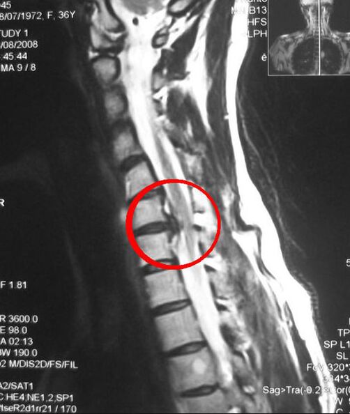
- Computed tomography (CT) and magnetic resonance imaging (MRI) are more accurate methods because they provide layer-by-layer images of the affected area.
- Electromyography is performed to differentiate neurological symptoms that develop due to nerve root compression in the thoracic type of osteoarthritis. Inspection is indicated if the following signs are present:
- impaired ability to coordinate movements;
- headache;
- dizzy;
- pressure fluctuations.
- Laboratory tests - performed to determine blood calcium levels and ESR (erythrocyte sedimentation rate).